
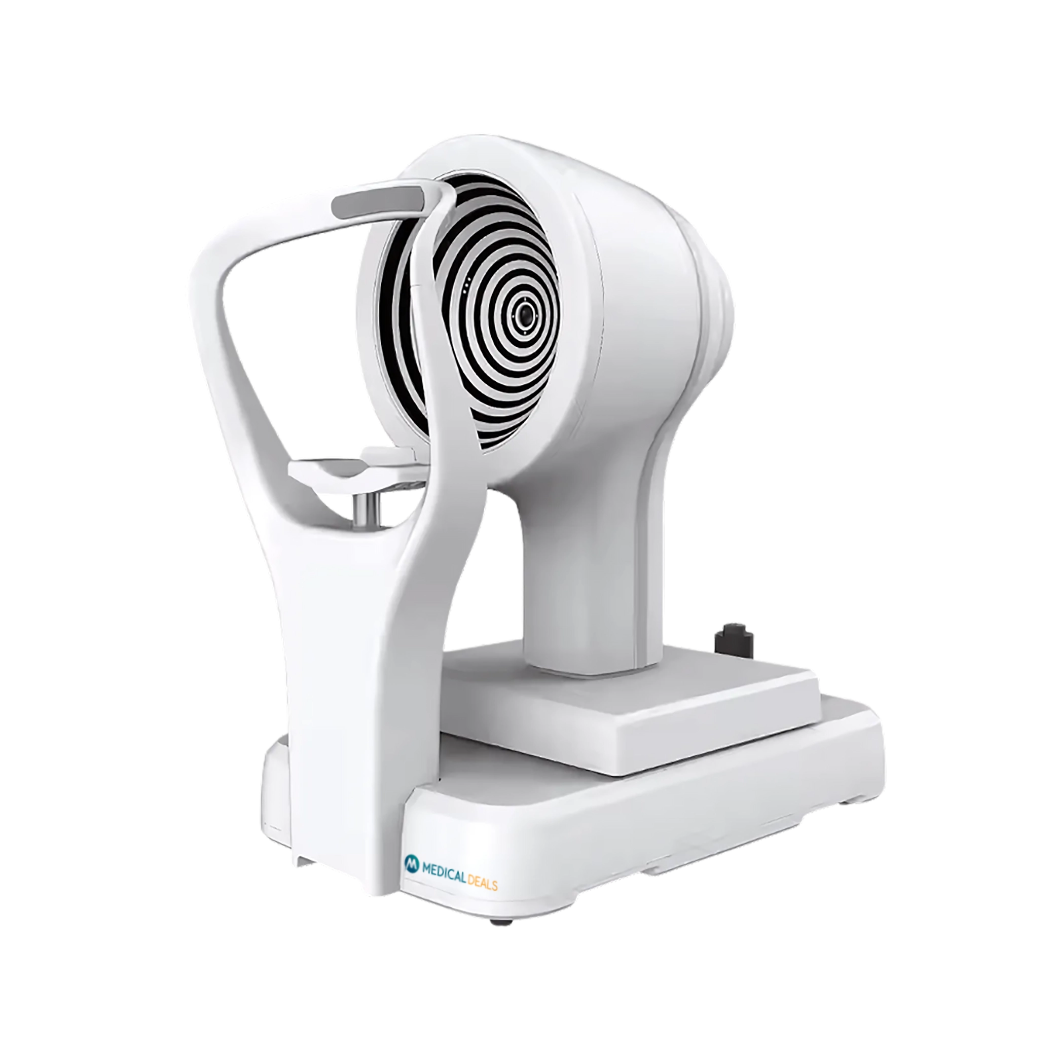
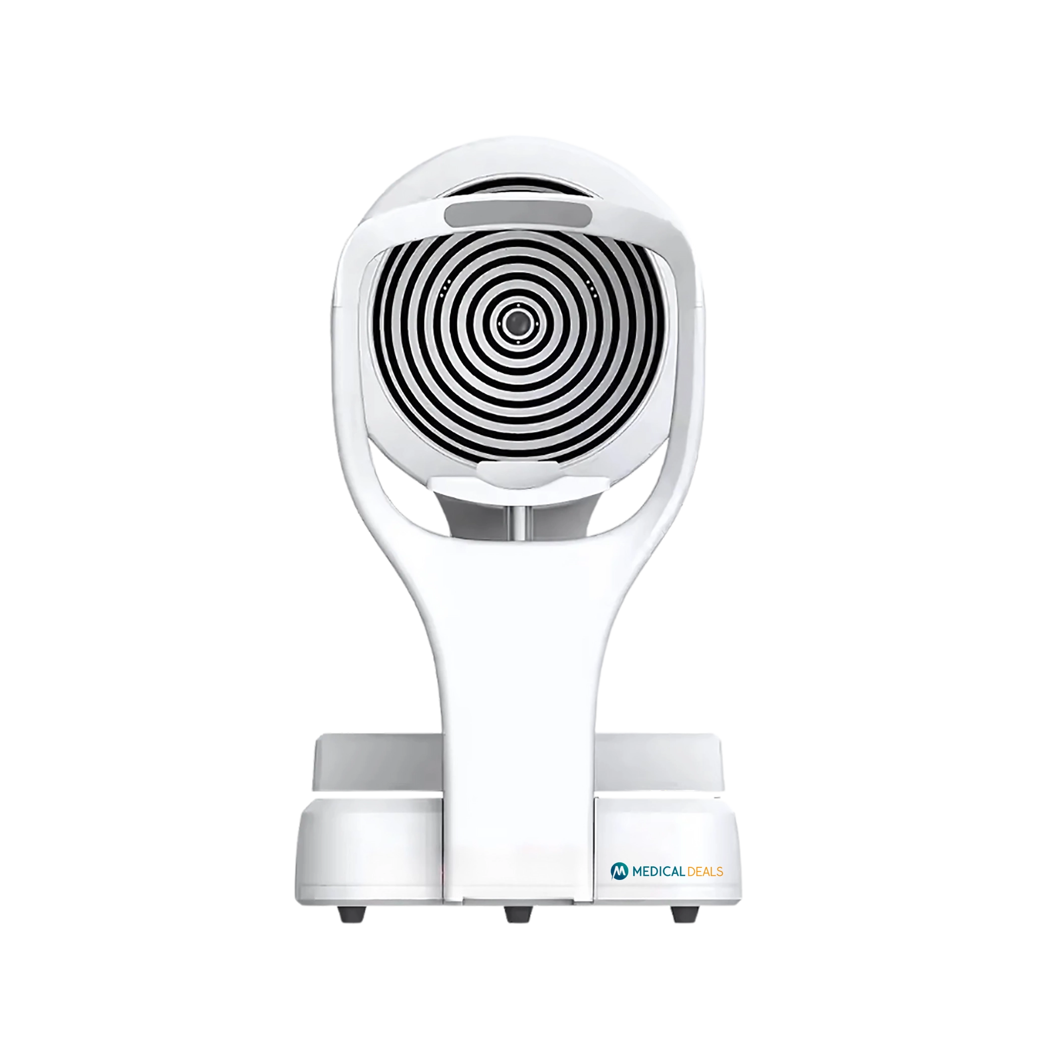
Ocular Osmolarity Analyzer MD-OOA-1001 operates with a near-infrared wavelength of 845 nm and blue light at 455 nm, ensuring high-accuracy imaging and analysis. It features a 2TB hard disk, Intel i7-8700 processor, and 16GB of memory, enabling seamless data processing and real-time image analysis. It analyzes eye closure dynamics to detect incomplete closure associated with ocular discomfort. Our product sets a new standard in dry eye diagnostics, providing clinicians with a reliable solution for comprehensive ocular evaluation.
Specifications
| Light source wavelength | Near infrared source: 845 nm Blue light: 455 nm |
| Computer system | Hard disk: Capacity 2T CPU: 17-8700 Graphic card: Intel 630(128MB/ASRROCK) Memory: 16 GB Monitor: AOC 241E1W/93 (23.8’’) |
| Imaging resolution | Colour camera: Less than 50 µm Near infrared enhanced camera: Less than 70 µm |
| Imaging modes | White light imaging modes Near infrared imaging modes Blue light imaging modes |
| Functions | Tear meniscus height measurement Analysis of tear film breakup time Lipid layer analysis Pinkeye analysis Analysis of eye closure Analysis of incomplete eye closure Meibomian gland detection Fluorescence image |
Features
Tri-Spectrum Imaging Technology
High-Resolution Optical System
AI-Enhanced Tear Film Analysis
Dynamic Fluorescence Visualization
Automated Tear Breakup Time Assessment
Ultra-Fast Image Processing Core
Dynamic Blink Analysis
Seamless EMR and Cloud Integration
Applications
Ocular Osmolarity Analyzer MD-OOA-1001 utilizes near-infrared hyperspectral imaging to reconstruct glandular morphology, quantifying lipid secretion kinetics and predicting progressive gland atrophy.
Medical Deals Ocular Osmolarity Analyzers offer high-resolution meibomian gland imaging, enabling detailed visualization of gland structures and functions. Their tear film lipid layer analyses provide real-time, high-definition video assessments, allowing clinicians to evaluate lipid thickness and distribution. Their non-invasive tear break-up time (NIBUT) analyses enable full corneal surface assessments without requiring fluorescein staining. Our solutions revolutionize dry eye management with seamless, user-friendly designs and unmatched clinical insights.

Address: 125 College Drive, Rock Springs,
WY 82901, USA
Email: info@medicaldeals.com
Phone: +44 7877 441082
WhatsApp: +447877441082